96 Hour Chick Embryo Serial Section
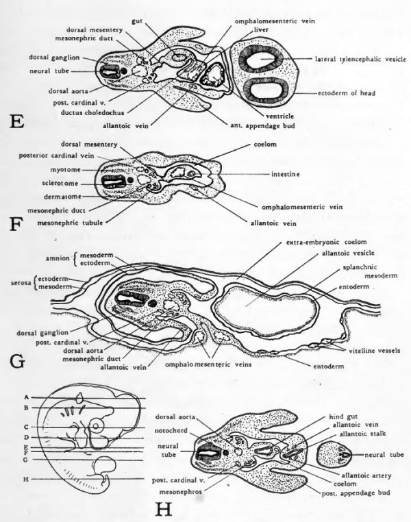
This membrane is made up of a bladderlike median ventral diverticulum of the hindgut endoderm, covered with splanchnic mesoderm. It connects with the hindgut, which will be found only in sections posterior to the posterior intestinal portal at this stage of development. Ultimately, this double membrane will fill the exocoel, and its outer layer of mesoderm will fuse with mesoderm of the chorion and the aminion and finally with the splanchnic mesoderm of the yolk sac splanchnopleure. Its function in the chick is related to respiration and excretion. The entire outer covering of the chick embryo is of ectodermal origin and is made up largely of squamous epithelium but will later also include horny scales, feather germs, quills and barbs, claws, beak coverings, and a temporary eggtooth. By evaginations from the surface, the linings of the following structures are also derived from ectoderm: the mouth (stomodeal portion and stomodeal hypophysis); cloaca (proctodeal portion); visceral clefts (peripheral halves); nostrils; eye chamber and lens; otic vesicles; and external auditory meatus.
Kak sostavitj kratkoe usloviya zadachi 3 klass. Kak-izbavitsjaot.ru is tracked by us since April, 2017.

Despite the relatively large size, the whole mounts (item# 311676) are cleared and stained. Serial sections (item# 311688) show the beginning of most of the. 48 hour chick serial sections. Crack keygen software. Lab practical. Thin layer of tissue surrounding the amnion. Exchanges gases and helps to provide oxygen to the embryo. In the chick this also brings calcium from the eggshell to the embryo in order to form the skeleton and beak. In mammals this forms part of the placenta. Derived from somatopleure.